Best Ultrasound Centre In Muzaffarnagar
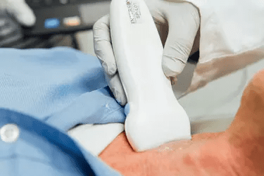
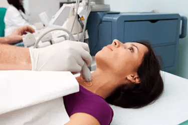
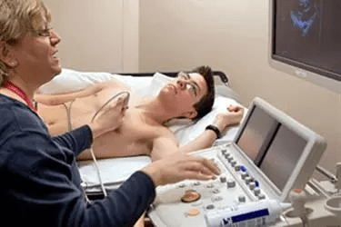
Ultrasound involves the use of high-frequency sound waves to create images of organs and systems within the body. An ultrasound machine creates images that allows various organs in the body to be examined. The machine sends out high-frequency sound waves, which reflect off body structures. The transducer has receptors which receive these signals and convert them into digital signals and form image of the body organ on the computer screen. Ultrasound is the gold standard for looking at the health of foetus in pregnant women. It allows the gynaecologists to detect any congenital abnormalties in the foetus and take corrective action.
4D Ultrasound
At Vardhman Hospital Muzaffarnagar we have the latest and most advanced 4-D Ultrasound machine. We have the latest 4-D Transvaginal and Transrectal Transducer which is extremely useful in diagnosing diseases of Uterus and Prostate. Transvaginal ultrasound is an internal ultrasound. It involves scanning with the ultrasound probe lying in the vagina or the rectum. Transvaginal ultrasound usually produces better and clearer images of the female pelvic organs, because the ultrasound probe lies closer to these structures. The female patients would usually prefer a female radiologist to perform the Transvaginal or TVS Studies and we have the female doctor for doing these studies at Vardhman Hospital.
Advantages Of 3D / 4D Ultrasound
3D/4D ultrasound can obtain views of the pelvis that are not seen on the conventional 2D ultrasound, especially views of the uterus. 2D ultrasound can obtain longitudinal and transverse views of the uterus. 3D ultrasound adds in the coronal view (or C-plane) of the uterus, enabling us to get an image of the uterus that is “front on”.
3D/4D ultrasound may help in the diagnosis and assessment of pelvic conditions, including:
- Congenital uterine abnormalities. Some women are born with changes in the shape of the uterus (for example, bicornuate uterus). Minor changes in the shape of the uterus are not uncommon.
- The location of endometrial polyps. The location of submucous fibroids and assessment of any intramural component.
- The location of an intrauterine contraceptive device (IUCD), especially if the IUCD is abnormally located (for example, penetrating into the wall of the uterus).
- Investigation of recurrent miscarriages and preterm deliveries.
Duplex Ultrasound In Muzaffarnagar
Pregnancy Ultrasound - In order to measure the growth of the foetus, ultrasound is the only diagnostic modality. During the nine months of the pregnancy at least 3 ultrasound exams are done, one in every trimester. In the first ultrasound of the pregnancy, the viability of the foetus is determined along with foetal heart rate and the gestational age. This ultrasound also gives the expected date of delivery.
During the second trimester around 14-15 weeks of pregnancy a very detailed and in-depth ultrasound is done called Level II Scan which gives complete information about the congenital abnormalities in the foetus. Not every radiologist is good at doing the Level 2 Scan and this is where highly experienced sinologists like Dr Singh come into the picture.
The cost of these ultrasounds are Rs. 1500 for the first Pregnancy Ultrasound done for foetal well being while the cost of Level 2 Ultrasound is Rs. 2500. The cost of Pregnancy Colour Doppler which is done in the 3rd Trimester just a few weeks before the expected delivery date is also Rs. 2500.


Types Of Ultrasounds
Ultrasound is a very versatile diagnostic modality and is widely used in the diagnosis of diseases in almost all parts of the body specially the diseases of the soft tissues. At Vardhman Hospital we perform the following Ultrasound Studies:
Breast ultrasound and Breast Elastography to study Breast Lumps and to find out whether they are benign or cancerous. The Breast Ultrasound is done in conjunction with Breast Mammography or MR Mammogram to study the Breast Lesions.
Abdominal ultrasound to study Liver, Gall Bladder, Pancreas, Spleen, Kidneys, Ureter plus Uterus and Ovaries in case of females. This ultrasound is done for any kind of abdominal pain to diagnose diseases like Appendicitis, Gall Bladder Stones, Ovarian Cyst, Uterine Fibroids, Kidney, Ureteric, and Urinary Bladder Stones, any kind of Abdominal Cancers. This is the cheapest but still very informative diagnostic test for doctors.
Scrotum Ultrasound is done to diagnose any problems in the testes like infection, inflammation, or undescended testis in case of children.
TVS or TRUS is the ultrasound where a thin probe is inserted in the vagina or the rectum to study the uterus, ovaries in females and prostate in males.
Small Part Ultrasound is the study of any part of the body like shoulder, neck, knee where the doctors are suspecting soft tissue injury like a ligament tear of fluid collection.
Ultrasound Guided Procedures are those in which ultrasound is used to provide guidance to the surgeon for doing FNAC, Biopsy, or to drain fluid collected inside the body where a very high level of precision is required.
Cost of Ultrasound Exams - Ultrasound is one of the cheapest diagnostic modality and is therefore the most popular. While most of the CT Scans cost between Rs. 2000-10000, most of the ultrasounds costs between Rs. 1000-2500 except some of the high-end color doppler studies involving multiple areas of the body. Since ultrasound exams do no involve any kind of radiation they can be performed on all people including children, old patients, and even pregnant women. Ultrasounds have zero side effects and therefore can be conducted any number of times in your entire life without causing any damage to the body. While CT Scan, MRI or even X-Rays are done by technicians, ultrasound can only be done by a trained doctor and therefore is not available at all places specially the government hospitals.
Patient Preparation
Most ultrasound procedures do not require advance preparation. Howsoever, there are certain exceptions which are listed as under:
Abdominal Ultrasound
Adults: Do not eat or drink 8 hours before the exam. Children: Do not eat or drink 4 hours before the study or skip 1 meal. Take medications with a small sip of water. If you are diabetic, please take your insulin
Pelvic Ultrasound Eat normally. One hour before your exam start drinking water. Do not empty your bladder until the exam is done
Bladder UltrasoundFor both male and female patients, start drinking water one hour before your exam. You must not urinate during this time so that the bladder can be filled with urine. Only if the bladder is full this study can be done.
Prostate-Transrectal UltrasoundIn this ultrasound the probe is inserted into your rectum after covering it with a condom. The rectum must be clean for this procedure to be done. Two hours before your exam, do a cleansing of the rectum with Enema.

