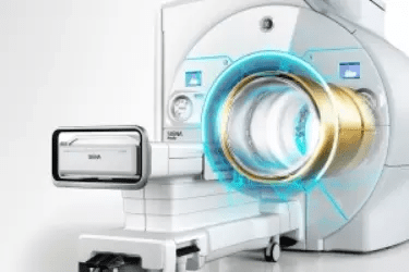
16 Channel MRI
Vardhman Hospital has installed the most advanced First 16 Channel MRI Machine in Muzaffarnagar. MRI is the gold standard of diagnostics in the field of brain, spine, joints, and abdomen. This is the last frontier in the field of radiology and there are few further investigations after this in diagnosing the disease. MRI works on the principle of getting information from the cells of each organ and therefore provides the most accurate information not only about the disease present in the body but also about the disease which may happen later in life. A good quality MRI machine differentiates between ordinary hospitals and super-specialty hospitals and Vardhman Hospital at Muzaffarnagar is the most advanced super specialty hospital in Western Uttar Pradesh.
Brain & Spine MRI
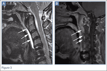
Comprehensive head and spine examinations can be performed with dedicated programs. . The Neuro Suite also includes protocols for diffusion imaging, perfusion Neurology imaging, and Functional MRI. The MRI of spine lets your doctor examine the small bones, called vertebrae, which make up your spinal column, as well as the spinal disks, spinal canal, and spinal cord. MRI scans of the spine are needed when conservative treatment is not working and more aggressive back pain treatments (e.g. spinal injections or surgery) are contemplated to relieve the symptoms.
MR Angiography
Excellent MR Angiography can be performed to visualize arteries and veins with or without a contrast agent. MRI Angiography is especially useful in identifying abnormalities such as aneurysms, blockage in carotid artery which can cause brain stroke, identifying arteries in the legs for endovascular surgery, very helpful in kidney transplant and diagnosing pulmonary embolism. The 16 Channel MRI at Vardhman Hospital is one of the best machines available in Muzaffarnagar for performing MR Angiography. The Non-Contrast MR Angiography is extremely useful in patients with deranged Kidney Function.
Breast & Pelivis MRI
MRI at Vardhman Hospital, Muzaffarnagar has dedicated breast coil for better and descriptive results. Cases of breast cancer are visualized better through MR Mammography. Also, MRI pelvis in cases of uterine fibroids is very useful in determining the size and the location of the fibroids enabling the gynecologist to plan the surgery for best outcomes. Excellent soft tissue differentiation, customized protocols (e.g. with fat saturation or water excitation or silicone excitation), as well as flexible multiplanar visualization, allow for fast, simple, and reproducible evaluation of MR breast examinations.
Patient Preparation For MRI
There’s no special preparation necessary for the MRI examination. Unless the staff at Vardhman Hospital specifically requests that you are not to eat or drink anything before the exam, there are no food or drink restrictions. You should continue to take any medication prescribed by your doctor unless otherwise directed. It is not allowed to wear anything metallic during the MRI examination at Vardhman Hospital, Muzaffarnagar, so it would be best to leave watches, jewelry, or anything made from metal at home. Even some cosmetics contain small amounts of metals, so it is best to not wear make-up. At Vardhman Hospital, we have a safe place to lock up valuables if you can’t leave them at home.
Now Digitizer Is Inside The Magnet
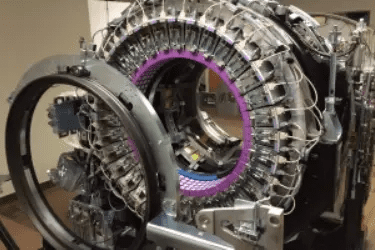
The earlier MRI machines required a high magnetic strength to get good quality images but the machine at Vardhman Hospital can get very good quality images with low magnetic strength due to a shift in the technology. The earlier machines were digitizing the data received from the patient in a digitizer which was located 12 feet from the patient due to the limitation of a strong magnetic field around the patient. This resulted in a loss of information but the MRI machine at Vardhman Hospital has the digitizer located just a few centimeters away from the patient and therefore there is no loss of information before it is digitized. In 2014 a new technology was developed whereby the digitizer after converting the analog signal to digital did not store it on a magnetic medium but on the optical cable.
Cardiac MRI
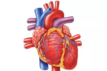
Cardiac MRI creates both still and moving pictures of your heart and major blood vessels. Doctors use cardiac MRI to get pictures of the beating heart and to look at its structure and function. These pictures can help them decide the best way to treat people who have heart problems. Cardiac MRI is also used for the patients who have kidney problems and cannot take contrast or dyes with iodine. Cardiac MRI is very common non invasive test for diagnosing damage caused to the heart muscle due to heart attack or pericarditis.
Whole Body MRI
The 16 Channel MRI at Vardhman Hospital, Muzaffarnagar, has a facility for Whole-Body MRI. Vardhman Hospital, Muzaffarnagar has 6 dedicated coils with an MRI machine to take care of the whole-body imaging. In the earlier MRI machines, the Signal to Noise Ratio was very high and therefore the doctors preferred CT Scan of the Abdomen over MRI but in the latest 360 degrees MRI this problem has been overcome, and now the MR Imaging of the abdomen has become the preferred diagnostic modality for GI Surgeons and Physicians
Precautions
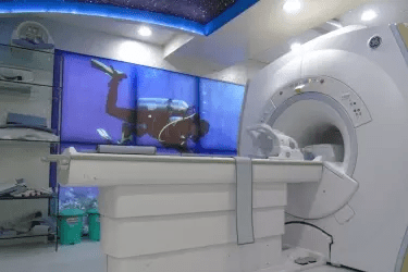
The patient will have to fill out a screening form asking about anything that might create a health risk or interfere with imaging. Below is a list of items that can create a health hazard during an MRI Scan.
- Pacemaker
- Implantable cardioverter-defibrillator (ICD)
- Neurostimulator
- Aneurysm clip
- Metal implant
- The implanted drug infusion device
- Foreign metal objects, especially if in or near the eye
- Shrapnel or bullet wounds
- Permanent cosmetics or tattoos
- Dentures/teeth with magnetic keepers
- Other implants that involve magnets
- Medication patch (i.e., transdermal patch) that contains a metal foil
